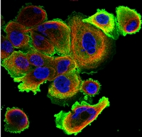
SCF (KITLG) Mouse Monoclonal Antibody
CAT#: PM1212P
SCF (KITLG) mouse monoclonal antibody, Azide Free
Need it in bulk or conjugated?
Get a free quote
CNY 3,630.00
Specifications
| Product Data | |
| Applications | ELISA, IF, WB |
| Recommend Dilution | Sandwich ELISA: In a Sandwich ELISA (assuming 100 µl/well), a concentration of 4.0-8.0 µg/ml of this antibody will detect at least 1000 pg/ml of recombinant human SCF when used with Biotinylated antigen affinity purified anti-Human SCF (Cat.-No PP1066B) as the detection antibody at a concentration of approximately 1.0-2.0 µg/ml. Western Blot: To detect Human SCF by Western Blot analysis this antibody can be used at a concentration of 0.25-0.50 µg/ml. Used in conjunction with compatible secondary reagents the detection limit for recombinant hSCF is 2.0-4.0 ng/lane, under non-reducing conditions. Immunohistochemistry: This antibody stained CACO-2 cells and A-431 cells. The primary antibody was incubated at 2.0 µg/ml overnight at 4°C followed by a fluorescent labeled secondary antibody. Information and photo are courtesy of the Cell Profiling group, SciLifeLab Stockholm. |
| Reactivity | Human |
| Host | Mouse |
| Clonality | Monoclonal |
| Immunogen | Highly purified (>98%) E.coli derived Recombinant Human SCF (Cat.-No PA124) |
| Specificity | Recognizes Human SCF. Other species not tested. |
| Formulation | PBS without preservatives State: Azide Free State: Lyophilized purified Ig fraction. |
| Reconstitution Method | Restore in sterile water to a concentration of 1.0 mg/ml. |
| Concentration | 1.0 mg/ml |
| Purification | Protein A Chromatography |
| Conjugation | Unconjugated |
| Storage Condition | Store lyophilized at 2-8°C for 6 months or at -20°C long term. After reconstitution store the antibody undiluted at 2-8°C for one month or (in aliquots) at -20°C long term. Avoid repeated freezing and thawing. |
| Gene Name | KIT ligand |
| Database Link | |
| Synonyms | KITL, Kit ligand, c-Kit ligand, Stem cell factor, Mast cell growth factor, MGF |
| Note | Protocol: Immunofluorescence: General Cell: 1. A multiwell plate (Glass bottom, 96-well, 300μL) is coated with fibronectin (conc. 12.5μg/mL) for 1 hour at room temperature (RT). |
| Reference Data | |
| Protein Families | Druggable Genome, Transmembrane |
| Protein Pathways | Cytokine-cytokine receptor interaction, Hematopoietic cell lineage, Melanogenesis, Pathways in cancer |
Documents
| Product Manuals |
| FAQs |
| SDS |
Resources
| 抗体相关资料 |


 United States
 Germany
 Japan
Japan
 United Kingdom
 China
China

